2020 Addled Egg Investigation
Lucy Knowles and Deborah Dawson, Department of Animal and Plant Sciences, University of Sheffield, UK
Introduction
A pair of Peregrines has nested on Leicester Cathedral for several years and webcams were set up on the nest to allow people around the world to view the breeding behaviour of these birds (leicesterperegrines.org.uk/streaming/). A new female Peregrine arrived at Leicester Cathedral in April 2020 and vicious fighting with the resident female took place. The incoming female took over the nest on April 27th and ousted the resident female. The resident female had laid four eggs in the nest box at Leicester Cathedral in March 2020, which were being incubated by the pair. The four eggs were laid on the 21st, 23rd, 26th and 28th March 2020 (Table 1, data taken from Leicester Peregrines Blog supplied by Jim Graham).
Incubation in Peregrines takes approximately 32–38 days and starts from the date the last egg was laid (Newton 1979). Locally, incubation times of 31–37 days have been recorded for the Peregrines nesting on St Georges Church in Sheffield (see www.sheffield.ac.uk/molecol/deborah-dawson/sheffieldperegrines, D. Dawson pers. comm, Table 4 of the webpage). In 2020, on the date the new female took over the nest in Leicester, the eggs had been incubated for 30 days and if fertile, were only 1–8 days from hatching (Table 1).
| Egg | Laying Date 2020 | Lay Time | Number of days until nest take-over* |
| 1 | 21st March | 05:30 | Failed: 37 days |
| 2 | 23rd March | 21:30 | Failed: 35 days |
| 3 | 26th March | 09:40 | Failed: 32 days |
| 4 | 28th March | 20:20 | Failed: 30 days |
*Number of days since the date the eggs were laid until the 27th April 2020 when a new incoming female drove away the resident female.
In Leicester, incubation dates from two previous clutches by the same pair in 2018 and 2019 were also 30 days for both years (Table 2). In 2018, four eggs were laid between 26th March and 4th April, with the first egg hatching on 5th May (only two eggs hatched). This gives 30 days incubation. In 2019, five eggs were laid between 20th March and 31st March, with the first hatching on 1st May (three of the five eggs hatched). This is also 30 days incubation from last laying until the first egg hatching, showing consistency over three years for Leicester Cathedral’s Peregrines.
| Egg | Laying Date 2018 | Lay Time | Hatch Date 2018 | Lay Time |
| 1 | 26th March | 17:30 | 5th May | 22:50 |
| 2 | 29th March | 15:15 | 7th May | 15:15 |
| 3 | 1st April | 16:00 | Failed | |
| 4 | 4th April | 06:50 | Failed |
| Egg | Laying Date 2019 | Lay Time | Hatch Date 2019 | Lay Time |
| 1 | 20th March | 09:00 | 1st May | 16:50 |
| 2 | 23rd March | 07:35 | 2nd May | 03:30 |
| 3 | 26th March | 01:45 | 3rd May | 07:40 |
| 4 | 28th March | 18:00 | Failed | |
| 5 | 31st March | 12:00 | Failed |
Incubation starts properly after the last egg is laid, which was the 28th March for 2020 at Leicester. Based on this, if fertile, the embryos at Leicester were expected to be at 30 days of development and were very close to hatching when incubation was disturbed/halted by the arrival of the new incoming female. Egg incubation became sporadic after the 27th April 2020.
The Peregrine nest on Leicester Cathedral was disturbed on the same day that it had been hoped the eggs may start hatching. One of the eggs in the nest was cracked open by the new incoming female and when she removed the contents, this egg was reported to contain feathers, so may have been due to hatch within 1–2 days. The new female also removed a second of the four eggs. The remaining two eggs were expected to hatch between 29th April and 7th May 2020 (based on 32–38 days from the date the last egg was laid (Newton 1979)).
Sadly, these remaining eggs did not hatch and in August were classed as no longer viable and removed from the nest by the Leicester Peregrine team on 13th August 2020. These addled eggs were stored in a fridge and passed on to the University of Sheffield to investigate if they were fertile and how much development had occurred. The plan was that if embryonic tissue was present, genomic DNA would be extracted to study Peregrine sex ratios and obtain a DNA profile of each individual. This would allow further study of the Leicester Cathedral Peregrine family and compare their genetic profiles with other Peregrines in the UK.
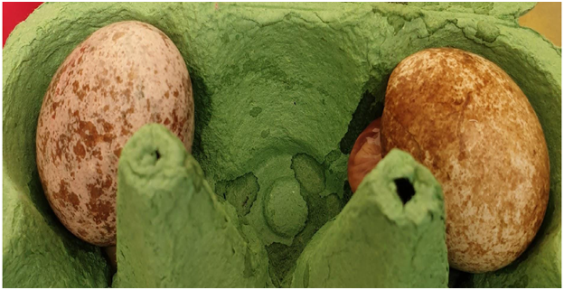
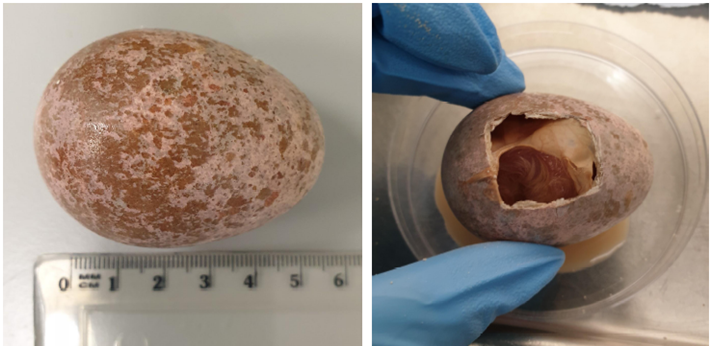
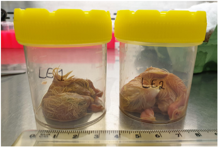
The egg at the right of the image (Egg 1, LE1) was brown and broken during transport from Bakewell to the University of Sheffield. Left egg was pink and remained intact (Egg 2, LE2).
Jim Graham of the Leicestershire and Rutland Ornithological Society delivered the two addled Peregrine eggs to Dr Douglas Ross at Bakewell Vet Clinic to be X-rayed on 4th September 2020. These were then passed on to Dr Deborah Dawson and Lucy Knowles of the University of Sheffield to investigate development and perform DNA analysis.
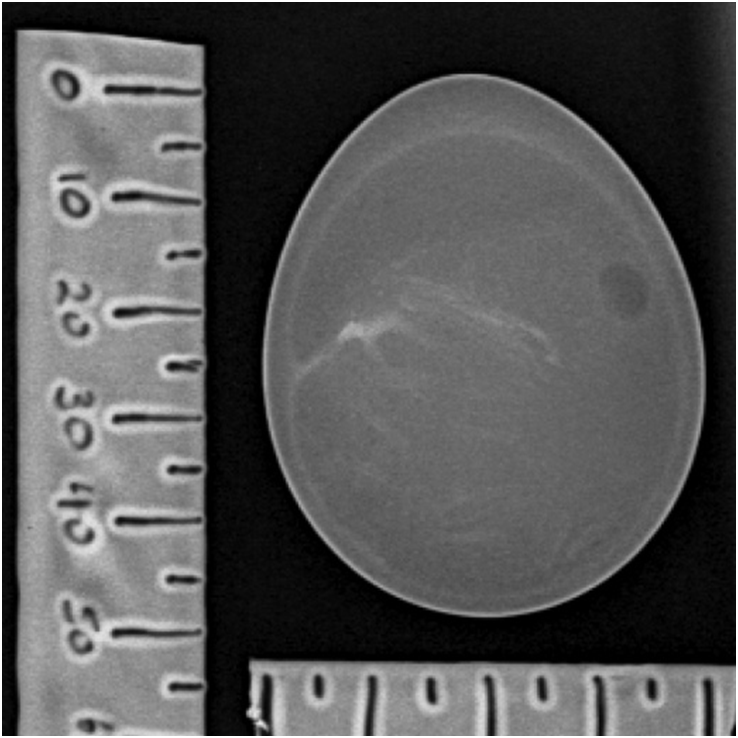
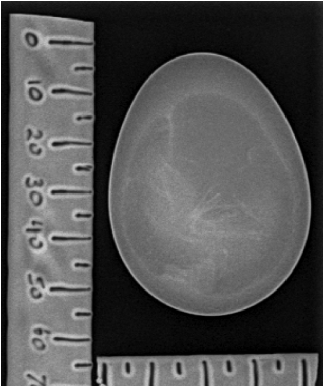
Figure 2: X-ray of the addled “brown” egg LE1, September 2020 (intact at this date). The X-ray displayed signs of skeletal development revealing the egg was fertile. The air space was small.
Figure 5: X-ray of the “pink” addled egg LE2. The X-ray of the pink egg also showed signs of skeletal development also confirming that the egg was fertile. There was a larger air space in this egg compared to the brown egg LE1.
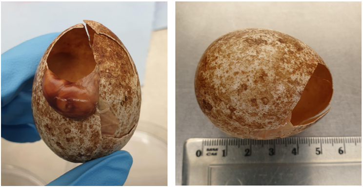
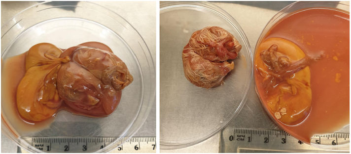
The brown egg (LE1) had a crack at the top that we suspect occurred during transit to the University of Sheffield from Bakewell on the day before it was opened on 18th September 2020, however the yolk had not broken (Figure 3). The hole in the eggshell was large enough to remove the unhatched chick. The embryonic sack containing the embryo was removed from the shell and the umbilical cord cut (Figure 3). The embryo had down covering most of its body and the beak and talons were clearly visible (Figure 4).
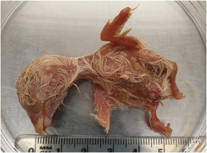
The embryo from the brown egg (LE1) was estimated to be close to hatching (N. Hemmings pers. comm.) and have had at least 26 days of development based on a comparison to development observed in chickens, scaled up based on the chickens, who have 21 days for full development prior to hatching (Hamburger & Hamilton 1951). Peregrines hatch after approximately 32-38 days after the date the last egg was laid (Newton 1979).
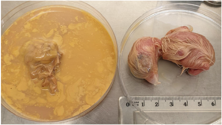
It was necessary to cut a larger hole because the first hole cut which was initially too small to remove the large chick. The yolk had broken (Figure 6). The chick was removed from the embryonic sack and the umbilical cord was cut.
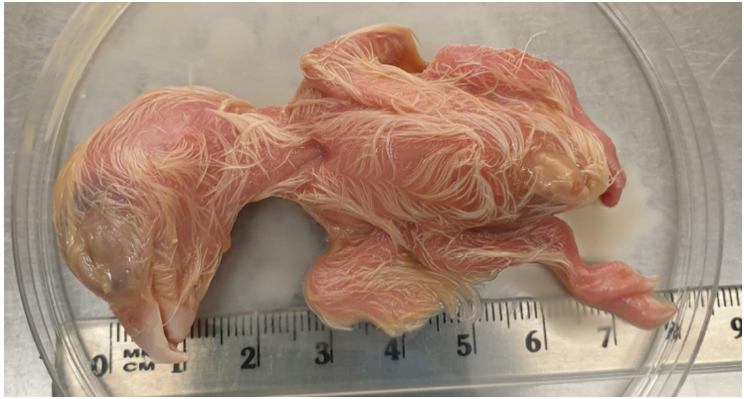
Embryo from the pink egg LE2 was 1-2 cm bigger than that from the brown egg LE1. Down covered almost all of the body and the beak and talons were clearly visible.
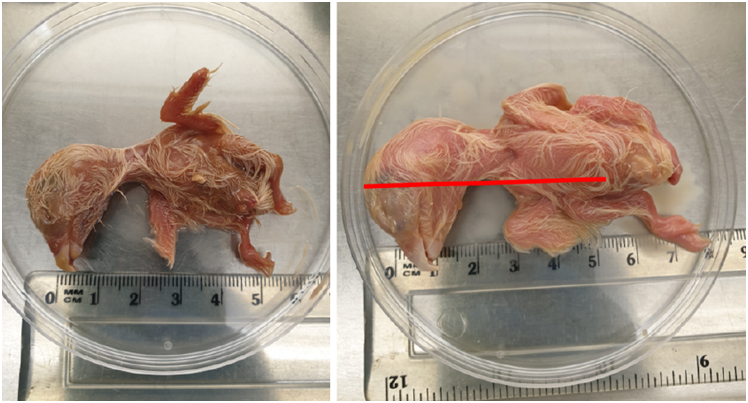
The red line represents the length of embryo LE1 on the image of embryo LE2 (Figure 9). An increase in length adds an increase in volume, hence embryo LE2 is a lot heavier than LE1.
Table 3: Comparison of development of the embryos from the brown egg LE1 and the pink egg LE2
| Embryo | Eggshell Colour | Embryo weight (g) | Observations | Estimated days incubated based on development |
| LE1 | Brown with speckles (cracked post X-ray) | 9.9 | Down covers most of the body, beak and claws clearly visible. Smaller air space. | 26 days |
| LE2 | Pink with speckles | 18.3 | Down covers almost all of the body, beak and claws clearly visible. Larger air space. | 30 days |
There is little data on embryo development in Peregrines. Chicken eggs hatch after approximately 21 days of incubation (Hamburger & Hamilton 1951). When scaling it up to Peregrines which hatch after 32-37 days of incubation (Newton 1979), this larger Peregrine embryo LE2 from the pink egg would be estimated to have ceased development at 30 days and the smaller embryo LE1 from a brown egg, a stopped developed a few days before this at approximately 26days.
The Leicester Peregrine’s Blog states that the male parent only incubated the two center eggs after/during the time when the mother and intruding female were fighting. The reduced development of the pink egg (embryo LE1) compared to the brown egg (embryo LE2) may be because the pink egg was located further from the centre of the nest. It is difficult to estimate the number of days incubated based on the embryo’s size/development since this has to be compared to embryo development in chicken (Hamburger & Hamilton 1951). It may be that both the brown egg and the pink egg were at the edge of the nest and incubation had both stopped development before the two centre eggs that were destroyed by the incoming female and not available to be studied. Since the last egg was laid on 28th March, the embryos should be at 30 days of development when the new female took over the nest 27th April and this agrees with the estimated stage of incubation based on the appearance, weight and size of the unhatched chicks for the larger unhatched chick (LE2).
As adults, female Peregrines are about a third bigger than males but at the hatching stage, chicks are similar sizes so this difference is not thought to be due to sex and genetic sexing revealed that both embryos are male (Table 4). In the last few days of development, the embryo grows rapidly (Hamburger & Hamilton 1951). The size difference suggests incubation of the brown egg (LE1) may have stopped 2-4 days before the pink egg (LE2) but both were very close to hatching.
DNA Sexing Results
Both embryos were sexed with three different sexing markers (Z43B, Z002E and P2D_P8; Dawson et al. 2016, Dawson 2007 and Dawson et al. 2009). The results of the three sex-typing markers were consistent for each embryo. Both embryos were identified as males (Table 4).
Table 4: DNA sexing of the two embryos from addled eggs
| Embryo | Egg colour | Z43B | Z002E | P2D_P8 | Sex |
| LE1 | Brown | 273, 273 | 117, 117 | 374, 374 | Male |
| LE2 | Pink | 273, 273 | 117, 117 | 374, 374 | Male |
DNA profiling and parentage of the Leicester Peregrines
Genomic DNA was successfully extracted from tissue taken from the embryos. A microsatellite DNA profile was obtained from both embryos which has been archived along with the DNA. This profile will be useful for future investigations of parentage at this nest and can be used to study and compare Peregrine populations. The DNA required for these future investigations can be obtained from chicks from mouth swabs or down feathers that are dislodged during ringing; or from adults via shed feathers or from embryos taken from addled eggs. We are also able to extract DNA and profile individuals from pellets if they have been collected and frozen soon after regurgitation.
There is some info about the parents of the embryos on the Leicester Peregrines Blog. The male parent of the chicks that hatched at Leicester prior 2020 (Table 2), and fathered the 2020 embryos, was ringed as a chick in Nottingham on 17th May 2018, making him 12 years old. We are not aware of any information on the resident female parent who was ousted in 2020. The incoming new female Peregrine at Leicester who turned up in Leicester in 2020 was ringed as a chick in Warwickshire on 30th June 2016 making her just four years old.
Acknowledgements
We thank Jim Graham of the Leicestershire and Rutland Ornithological Society for providing the two addled Peregrine eggs for analysis and nest data. Dr Douglas Ross of Bakewell Veterinary Clinic kindly took X-rays of the eggs. We thank Dr Nicola Hemmings of the University of Sheffield for viewing the photos, providing expert advice and assessing the stage of development for the two embryos.
References
Dawson DA (2007); Genomic analysis of passerine birds using conserved microsatellite loci. PhD Thesis, University of Sheffield, UK.
Dawson DA, dos Remedios N, Horsburgh GJ (2016); A new marker based on the avian spindlin gene that is able to sex most birds, including species problematic to sex with CHD markers. Zoo Biology, 35, 533–545. [Open Access]
Dawson DA, Horsburgh GJ, Krupa AP, Stewart IRK, Skjelseth S, Jensen H, Ball AD, Spurgin LG, Mannarelli M-E, Nakagawa S , Schroeder J et al (2012); Microsatellite resources for Passeridae species: a predicted microsatellite map of the house sparrow Passer domesticus. Molecular Ecology Resources, 12(3), 501–523.
Hamburger V & Hamilton HL (1951) A Series of Normal Stages in the Development of the Chick Embryo. Journal of Morphology, Jan; 88(1):49–92.
Leicester Peregrines Blog: leicesterperegrines.org.uk/
Newton I (1979) Population Ecology of Raptors. T & AD Poyser Ltd. Table 18, p343.
Sheffield Peregrines Blog: sheffieldperegrines.wordpress.com
Sheffield Peregrines DNA Study:
www.sheffield.ac.uk/molecol/deborah-dawson/sheffieldperegrines
Lucy Knowles and Deborah Dawson,
Department of Animal and Plant Sciences
University of Sheffield, UK
NB: As an educational tool, it is hoped that the chicks will be stuffed and shown in a nest box as part of a display in the Alfred Denny museum at Sheffield University. It is also proposed that the skeletons are assembled as part of the display.
The tissue samples will be banked the -80°C freezer for reference and future DNA studies/investigations.